持向量机与独立分量分析在MR脑图像分割中的应用研究

VIP免费
3.0 陈辉 2024-11-19 5 4 1.1MB 80 页 15积分
侵权投诉
I
摘 要
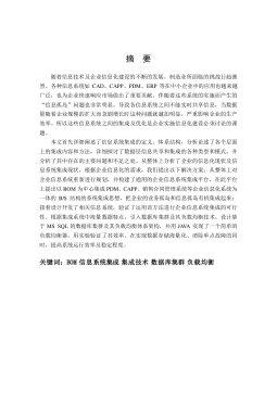
医学图像分割是医学图像可视化、人体组织定量测量等临床应用的先决条件。
尽管目前有很多的医学图像分割方法,但由于医学图像的复杂性及对分割精度的
特殊要求,目前还没有一种方法能有效地解决医学图像分割问题。临床应用中主
要以人工分割和半自动分割方法为主。人工分割方法耗时费力,且分割结果难于
再现;半自动分割方法把操作者的知识和计算机数据处理能力有机地结合起来,
从而实现对医学图像的半自动(即交互式)分割。与人工分割方法相比,半自动
分割方法大大减小了人为因素的影响,而且分割速度更快,但操作者的知识和经
验仍然是影响图像分割质量的重要因素。全自动分割方法充分利用计算机数据处
理能力,完全摆脱人为因素的影响,分割速度快,分割结果再现性好,但分割质
量取决于所使用的分割算法。能实现对医学图像快速、精确的全自动分割,一直
是人们追求的目标。在众多的全自动分割方法中,基于机器学习分类技术的分割
方法一直是人们研究的热点。而基于VC维和统计学习理论的支持向量机Support
Vector MachinesSVM)方法由于其数学形式简单、全局寻优、学习速度快、
化能力优、适合处理高维数据等特点而被广泛应用于图像分割中。
SVM在使用结构风险最小化原则替代经验风险最小化原则的基础上,综合了
统计学习和神经网络等方面的技术,并被证明可在最小化结构风险的同时,有效
地提高算法的推广能力。但SVM本质上是解决两类分类问题的,而实际应用中常
常需要解决多类分类问题,MR脑组织分割问题。基于这一实际应用,本文研究
了一种解决多分类问题的SVM方法,可以一次性把MR脑图像中的脑白质、脑灰质
及脑脊液等不同脑组织分别提取出来。另外,SVM中不同的核函数及相应模型参
数对其分类性能有重要影响,本文研究了针对MR脑组织分割问题的核函数及模型
参数选择问题,从中选取最优核函数及模型参数进行MR脑图像分割。
脑组织分割实质上是对脑组织的分类问题。在分类问题中,分类性能的好坏
还取决于对分类特征的选择。不同的特征对分类性能有不同的贡献。过多的特征
可能导致过学习,降低分类器泛化性能,同时分类特征维数过高可能导致“维数
灾”问题,增加分类器算法复杂度,降低分类器性能。特征不足又不足以描述数
据空间,影响分类的质量。因此,在分类过程中对所选择的特征进行优化处理对
提高分类速度和质量至关重要。在现有的MR脑图像分割研究中,一般都是选取单
一灰度特征进行分类研究,仅有个别文献采用多特征进行脑图像分割,但这些文
献中并未对选取的特征进行优化处理,由于各特征间可能有相关性,存在冗余信
II
息,从而影响了分割质量和分割速度。
独立分量分析Independent Component AnalysisICA方法是在盲源分离问
题上发展起来的一种新的多维信号处理方法,能有效地从源信号中提取出独立信
息,是一种有效的冗余信息消除技术。本文详细研究了独立分量分析理论及实现
技术,并根据MR脑图像分割问题,引入ICA对所提取的图像特征进行优化处理,
消除特征相关性,降低原始特征维数,提高SVM分类器分类性能。
本文通过对SVMICA相关理论的研究,提出了结合SVMICA两种方法解决
MR脑图像分割问题。研究结果表明:独立分量分析是一种有效的多维数据处理方
法,成功地去除了原始图像特征的冗余信息,降低特征维数,提高了SVM分类性
能;基于SVMICAMR脑图像分割方法成功地结合了各自的优点,取得了比单
一方法更好的分割结果。
关键词:医学图像分割 支持向量机 独立分量分析 磁共振脑图像
III
ABSTRACT
Medical image segmentation is prerequisite to medical image visualization and
tissue measurement. There are many medical image segmentation methodsbecause
of the complexity of medical image and clinical special requirement for the
segmentation precisionnone of these methods have successfully settled the problem
of medical image segmentation so far. Clinical application relies mainly on manual
segmentation and semi-automatic segmentation. Manual segmentation is
time-consuming and laborious and its segmentation is hard to be duplicated;
semi-automatic segmentation integrates radiologists’ knowledge with data processing
capability of computer. Compared with manual segmentation semi-automatic
segmentation greatly reduces the effects of human and is faster but radiologists’
knowledge and experience will still affect the quality of medical image segmentation.
Auto-segmentation makes full use of the data processing capability of computer and
entirely gets rid of influence of human being the segmentation is faster and
duplicated easier but the segmentation equality is based on the segmentation
algorithm. Fast and precise medical image segmentation is always the pursuit goal of
researchers. Among the existing auto-segmentation methods classification
technology based on machine learning is the highlight of current image segmentation
research. Due to its simple mathematical expression global optimization fast
learning ratehigh generalization performance and suitable for high dimension data
processing support vector machine (SVM) based on Vapnik-Chervonenkis
Dimension and statistical learning theory is widely used in image segmentation.
Based on the principle of structural risk minimization instead of principle of
experiential risk minimizationand combining the techniques of statistical learning
machines learning and neural networks etcit is proved that SVM has good capability
of generalization with minimization of experiential risk. Essentially SVM is a
technique for solving two-class classification problem but in practical application
multi-class classification problem was taken into consideration commonlysuch as
Magnetic Resonance Imaging (MRI) segmentation. So this paper studied a multi-class
SVM algorithm to segment gray matter (GM)white matter (WM) and cerebrospinal
IV
fluid (CSF) from MRI at one time. Different kernel functions and model parameters
have different contributions to SVM classificationthis paper studied the selection
problems of kernel functions and model parameters in order to obtain optimal kernel
function and model parameters for MRI segmentation.
Brain tissues segmentation is actually a classification problem for brain tissue.
In classification problemclassification performance is also based on the selection of
classification features. Different features have different contribution to classification
performanceto select too much features may result in over-learning and degrade the
generalization performanceand too much features also may lead to “dimension risk”
problem and increase the complexity of classification algorithm. To select too few
features will not describe the data space correctly. So features optimization in
classification is very important to speed the classification and increase the
classification equality. In the most current MRI segmentation research literatures only
gray feature was commonly used for brain tissues classification a few research
papers apply multi-features to MRI segmentationbut the selected features did not be
optimized. Because between various features may exist the relevance and redundant
information the segmentation quality and speed may be affected seriously if the
selected features without optimized.
Independent Component Analysis (ICA) is a multi-dimension signal processing
method developed on the basis of blind source separation; it’s efficient to separate
independent information out of source signal and eliminate redundant information.
According to the practical application of MRI segmentationICA is introduced to
process the selected image featureseliminate the relevance between different features
and reduce the feature dimensions to improve the classification performance.
Trough the study of SVM and ICA theorythis paper presented a method to
solve MRI segmentation problem by combining SVM and ICA. Experiment results
have showed that ICA is an efficient multi-dimension signal processing method; it can
successfully eliminated redundant information from original features reduced the
number of feature dimension and improved the performance of SVM classifier. The
classification method based on combining SVM and ICA can get better segmentation
result of brain tissues than using single method.
Key Word: Medical image segmentationSupport vector machines
Independent component analysisMagnetic resonance image
V
目录
............................................................... I
ABSTRACT ...........................................................III
目录 ................................................................. V
第一章 绪论 .......................................................... 1
§1.1 引言 ........................................................ 1
§1.2 医学图像分割方法概述 ....................................... 4
§1.3 机器学习分类方法综述 ....................................... 5
§1.3.1 传统学习分类方法 ...................................... 5
§1.3.2 传统学习分类方法存在的问题 ............................ 6
§1.3.3 统计学习理论与支持向量机方法 .......................... 7
§1.4 独立分量分析概述 ........................................... 8
§1.5 本文主要研究工作 ........................................... 9
§1.6 本文结构 .................................................. 10
第二章 医学图像分割方法综述 ......................................... 12
§2.1 引言 ...................................................... 12
§2.2 MR 图像分割的目的和意义 .................................... 12
§2.3 MR 图像分割方法研究综述 .................................... 13
§2.4 医学图像分割方法的评价 ................................... 18
§2.5 本章小结 ................................................... 19
第三章 基于支持向量机的 MR 脑图像分割研究 ............................ 21
§3.1 引言 ...................................................... 21
§3.2 统计学习理论 .............................................. 22
§3.2.1 机器学习问题的描述 ................................... 22
§3.2.2 经验风险最小化 ....................................... 22
§3.2.3 学习机器的 VC 维 ...................................... 23
§3.2.4 推广性的界 ........................................... 23
§3.2.5 结构风险最小化 ....................................... 24
§3.3 支持向量机原理 ............................................ 25
§3.3.1 线性可分的最优分类面 ................................. 26
§3.3.2 线性不可分的最优分类面 ............................... 28
§3.3.3 支持向量机 ........................................... 29
§3.3.4 支持向量机实现算法 ................................... 30
§3.3.5 支持向量机学习算法的步骤 ............................. 33
§3.4 多类支持向量机 ............................................ 33
§3.5 支持向量机方法特点 ........................................ 34
§3.6 基于支持向量机的 MR 脑图像分割 ............................. 35
§3.6.1 图像预处理 ........................................... 36
§3.6.2 图像特征提取 ......................................... 37
VI
§3.6.3 训练与测试样本选取 ................................... 41
§3.6.4 图像分割实验 ......................................... 42
§3.7 本章小节 .................................................. 46
第四章 独立分量分析及其在图像处理中的应用研究 ....................... 48
§4.1 引言 ...................................................... 48
§4.2 独立分量分析的数学描述 .................................... 48
§4.2.1 独立分量分析的约束限制条件 ........................... 49
§4.2.2 ICA 独立性度量 ....................................... 50
§4.3 独立分量分析算法研究 ...................................... 53
§4.3.1 独立分量分析的实现算法 ............................... 53
§4.3.2 独立分量分析分离性能的两个衡量指标 ................... 56
§4.4 基于独立分量分析的混合图像分离 ............................. 56
§4.5 本章小结 .................................................. 59
第五章 基于支持向量机及独立分量分析的 MR 脑图像分割 ................. 60
§5.1 引言 ...................................................... 60
§5.2 基于独立分量分析的图像特征提取 ............................ 61
§5.3 基于支持向量机及独立分量分析的 MR 脑图像分割 ................ 61
§5.4 本章小结 .................................................. 67
第六章 总结与展望 .................................................. 68
§6.1 本文所做研究工作及创新点 .................................. 68
§6.2 今后进一步的研究方向 ...................................... 69
参考文献 ............................................................ 70
在读期间公开发表的论文和承担科研项目及取得成果 ...................... 75
.............................................................. 76
摘要:

I摘要医学图像分割是医学图像可视化、人体组织定量测量等临床应用的先决条件。尽管目前有很多的医学图像分割方法,但由于医学图像的复杂性及对分割精度的特殊要求,目前还没有一种方法能有效地解决医学图像分割问题。临床应用中主要以人工分割和半自动分割方法为主。人工分割方法耗时费力,且分割结果难于再现;半自动分割方法把操作者的知识和计算机数据处理能力有机地结合起来,从而实现对医学图像的半自动(即交互式)分割。与人工分割方法相比,半自动分割方法大大减小了人为因素的影响,而且分割速度更快,但操作者的知识和经验仍然是影响图像分割质量的重要因素。全自动分割方法充分利用计算机数据处理能力,完全摆脱人为因素的影响,分割...

展开>> 收起<<
持向量机与独立分量分析在MR脑图像分割中的应用研究.pdf

共80页,预览8页

还剩页未读, 继续阅读

作者:陈辉 分类:高等教育资料 价格:15积分 属性:80 页 大小:1.1MB 格式:PDF 时间:2024-11-19

开通VIP享超值会员特权

  • 多端同步记录
  • 高速下载文档
  • 免费文档工具
  • 分享文档赚钱
  • 每日登录抽奖
  • 优质衍生服务
/ 80
客服
关注